Breast Cancer Screening & Genetics
Your Body is Your Temple
Research, Resources & Education
Table of Contents
Breast cancer is one of the most pervasive cancers affecting women worldwide. Unfortunately, the affliction does not discriminate based on age, race, or socioeconomic status. Therefore, it is crucial that women prioritize their breast health by getting regular screenings. If one is found to be at higher risk, genetic testing can provide more personalized care options. Genetic testing would identify if any mutations are present that increase the chances of a patient developing breast cancer.
Information and pictures from National Cancer Institute unless otherwise specified
Types of Screenings Video
YouTube Videos that help explain different ways of screening for breast cancer.
Disclaimer:
This is for research only and Lost Temple Fitness & Cancer does not endorse any video presented on this website.
It is advised that you ALWAYS CHECK WITH YOUR PHYSICIAN for a proper diagnosis and treatment plan.
Breast Cancer Screening
6 Videos
Screenings
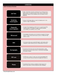
Lab Tests
High or low levels of certain substances in your body can be a sign of cancer. So, lab tests of your blood, urine, or other body fluids that measure these substances can help doctors make a diagnosis.
- Some lab tests involve testing blood or tissue samples for tumor markers.
- Tumor markers are substances that are produced by cancer cells or by other cells of the body in response to cancer.
- Most tumor markers are made by normal cells and cancer cells but are produced at much higher levels by cancer cells
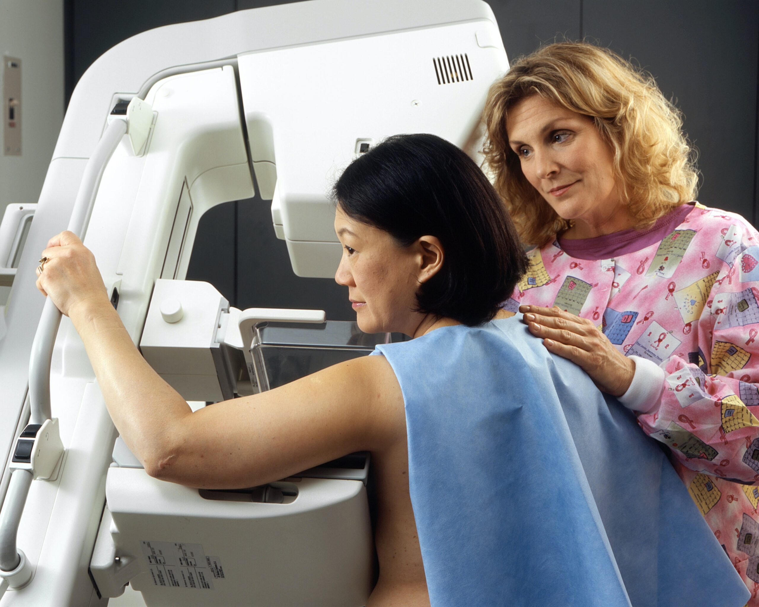
Screening Mammogram
X-rays of the breasts taken to check for breast cancer in the absence of signs or symptoms.
Possible mammogram findings include:
- Lumps (mass or tumor). Lumps come in different sizes and shapes.
- Fluid-filled cysts are usually smooth and rounded, with clear, defined edges and are not cancer.
- Lumps that have a jagged outline and an irregular shape are of more concern.
- Calcifications. There are two types of breast calcifications, or calcium deposits:
- Macrocalcifications, which look like small white dots on a mammogram.
- They are often caused by aging, an old injury, or inflammation and are usually benign.
- Microcalcifications, which look like white specks on a mammogram.
- If found in an area of rapidly dividing cells or grouped together in a certain way, they may be a sign of DCIS or breast cancer.
- Macrocalcifications, which look like small white dots on a mammogram.
- Dense breast tissue: A dense breast has relatively less fat and more glandular and connective tissue. This mammogram finding is both common and normal, especially among younger women and women who use menopausal hormone therapy.
- Dense breast tissue can make a mammogram more difficult to interpret because both dense breast tissue and breast tumors appear as solid white areas in the image
Diagnostic Mammography
A type of mammography in which more x-ray pictures of the breast are taken from different angles to allow a possible abnormality to be examined more closely.
- X-ray of the breasts used to check for breast cancer after a lump or other sign or symptom of breast cancer has been found.
Ultrasound
A procedure that makes a picture (called a sonogram) of breast tissue in order to find out if a lump is solid or is filled with fluid (that is, a cyst).
- Pictures are made using sound waves.
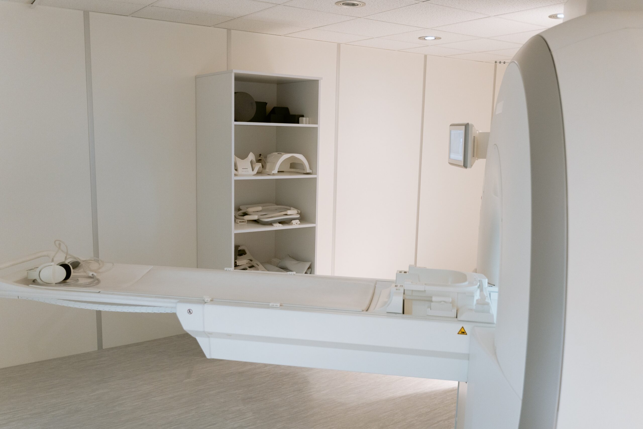
Magnetic Resonance Imaging - MRI
A procedure that uses a powerful magnet, radio waves, and a computer to take detailed pictures of areas inside the breast.
- An MRI can be used to learn more about breast lumps or large lymph nodes that were found during a clinical breast exam or breast self-exam but were not seen on a mammogram or ultrasound.
Ductography - AKA Galactography
A procedure that takes pictures of the breast ducts, so that doctors can learn more about certain kinds of abnormal nipple discharge or masses such as intraductal papillomas (wart-like tumors that are benign).
- Pictures of the breast ducts are taken using a contrast material that is given through an injection to help breast ducts show up clearly.
PET scan
A PET scan is a type of nuclear scan that makes detailed 3-D pictures of areas inside your body where glucose is taken up.
- Because cancer cells often take up more glucose than healthy cells, the pictures can be used to find cancer in the body.
- Before the scan, you receive an injection of a tracer called radioactive glucose.
- During the scan, you will lie still on a table that moves back and forth through a scanner.
Biopsy
A procedure that removes a sample of breast tissue or an entire lump so that it can be checked for signs of disease.
- Imaging procedures (such as ultrasound, MRIs, or x-rays) are often used during a biopsy to guide the surgeon.
- A pathologist then examines the sample under a microscope or performs other tests on it.
Common types of breast biopsies include:
- Core needle biopsy: The use of a wide needle to remove small tissue sample(s) that are about the size of a grain of rice. It may cause a temporary bruise. Also called core biopsy.
- Fine-needle aspiration biopsy: The use of a thin needle to drain fluid and/or to remove cells.
- Surgical biopsy: The removal of part, or all, of a lump so it can be checked for signs of cancer.
- An incisional biopsy removes a sample of breast tissue.
- An excisional biopsy removes an entire lump or suspicious area.
- Wire localization (also called needle localization and needle (wire) localization) may be used to mark the area of abnormal tissue before the biopsy.
- Vacuum-assisted biopsy: The removal of a small sample of breast tissue using a probe that is connected to a vacuum device.
- The small cut made in the breast is much smaller than with surgical biopsy. This procedure causes little scarring, and no stitches are needed. It may also be called vacuum-assisted core biopsy.
A biopsy procedure that uses a computer and a 3-dimensional scanning device to find a tumor site and guide the removal of tissue for examination under a microscope.
- Biopsies are usually done in a doctor’s office or a clinic on an outpatient basis.
- This means you will go home the same day as the procedure.
- Local anesthesia is used for many biopsies, so you’ll be awake but won’t feel pain during the procedure.
- General anesthesia is commonly used for surgical biopsies, which means you’ll be asleep during the procedure.
Genetics & Cancer Videos
YouTube Videos that help explain different genes that can increase your chance of breast cancer.
Disclaimer:
This is for research only and Lost Temple Fitness & Cancer does not endorse any video presented on this website.
It is advised that you ALWAYS CHECK WITH YOUR PHYSICIAN for a proper diagnosis and treatment plan.
Genetics
11 Videos
Genetics & Cancer
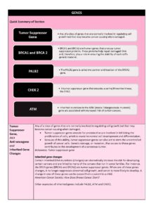
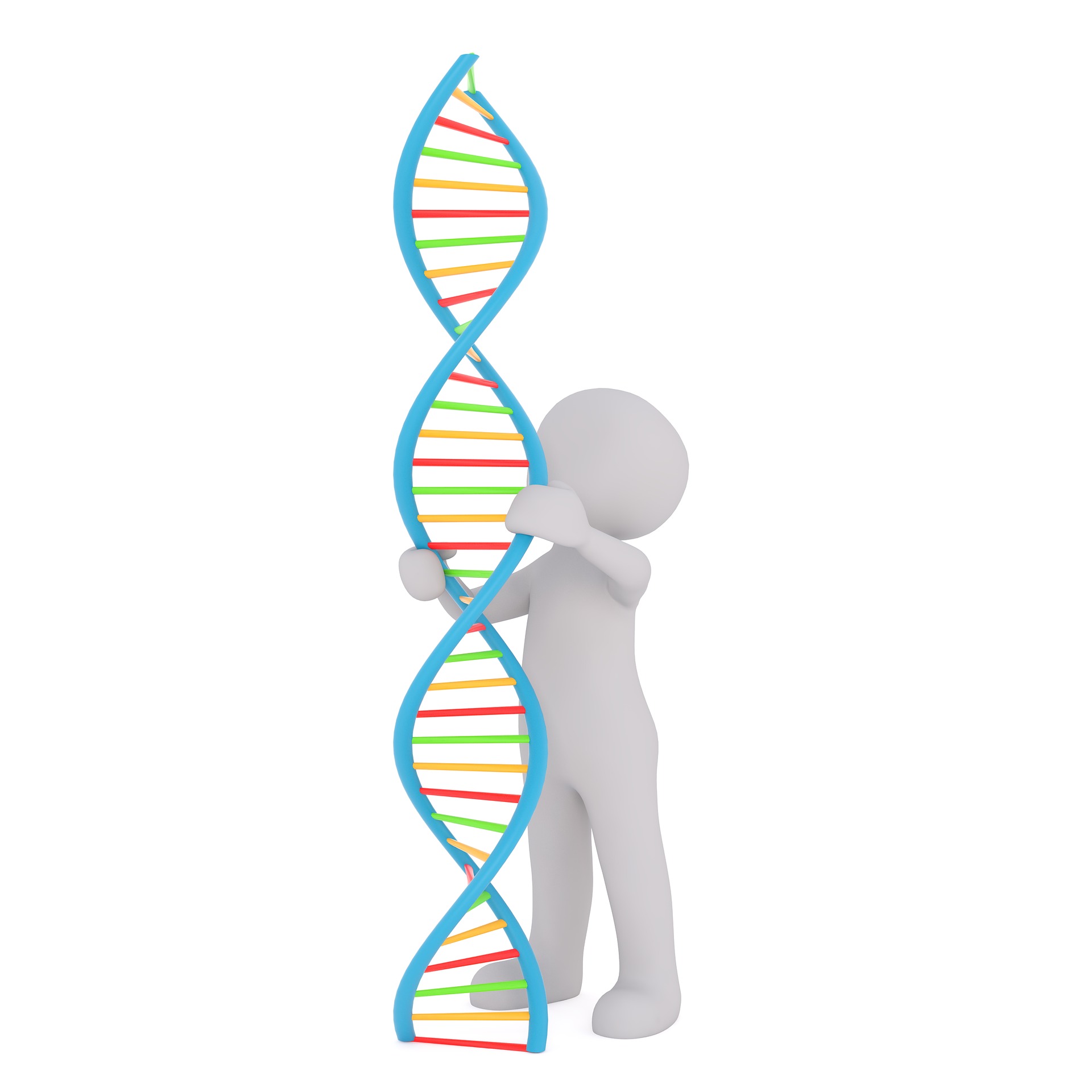
Tumor Suppressor Gene, AKA Anti-oncogene or Inherited Gene Changes
Any of a class of genes that are normally involved in regulating cell growth but that may become cancer-causing when damaged.
- Tumor suppressor genes encode for proteins that are involved in inhibiting the proliferation of cells, which is crucial to normal cell development and differentiation.
- Because of this ability, tumor suppressor genes can also act to stem the uncontrolled growth of cancer cells. Genetic damage, or mutation, that occurs to these genes contributes to the development of a cancerous tumor. (Britannica: Tumor suppressor gene)
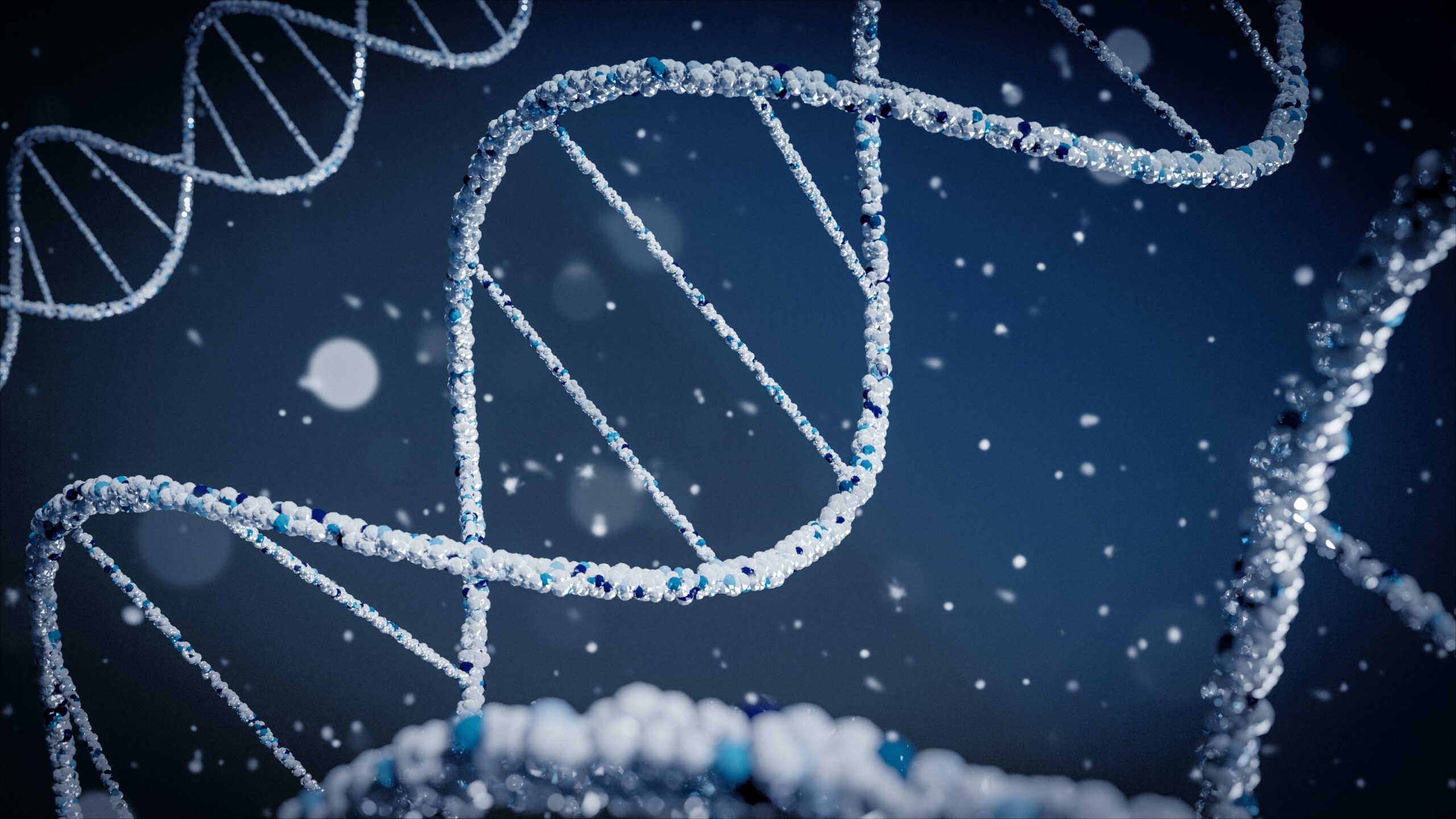
Inherited gene changes
Certain inherited DNA mutations (changes) can dramatically increase the risk for developing certain cancers and are linked to many of the cancers that run in some families.
- For instance, the BRCA genes (BRCA1 and BRCA2) are tumor suppressor genes. When one of these genes changes, it no longer suppresses abnormal cell growth, and cancer is more likely to develop.
- A change in one of these genes can be passed from a parent to a child. (American Cancer Society: What Causes Breast Cancer?)
Other examples of inherited genes include PALB2, ATM and CHEK2.
BRCA1 and BRCA 2
BRCA1 and BRCA2 are human genes that produce tumor suppressor proteins. These proteins help repair damaged DNA and, therefore, play a role in ensuring the stability of each cell’s genetic material.
- When either of these genes is mutated, or altered, such that its protein product is not made or does not function correctly, DNA damage may not be repaired properly.
- As a result, cells are more likely to develop additional genetic alterations that can lead to cancer.
- Specific inherited mutations in BRCA1 and BRCA2 most notably increase the risk of female breast and ovarian cancers, but they have also been associated with increased risks of several additional types of cancer.
- People who have inherited mutations in BRCA1 and BRCA2 tend to develop breast and ovarian cancers at younger ages than people who do not have these mutations.
- A harmful BRCA1 or BRCA2 mutation can be inherited from a person’s mother or father. Each child of a parent who carries a mutation in one of these genes has a 50% chance (or 1 chance in 2) of inheriting the mutation.
- About 12% of women in the general population will develop breast cancer sometime during their lives.
- By contrast, a recent large study estimated that about 72% of women who inherit a harmful BRCA1 mutation and about 69% of women who inherit a harmful BRCA2 mutation will develop breast cancer by the age of 80.
- There also is a higher risk of developing a new primary cancer in the opposite (contralateral) breast in the years following a breast cancer diagnosis.
Who should get tested: Several screening tools are available to help health care providers with this evaluation:
- Breast cancer diagnosed before age 50 years
- Cancer in both breasts in the same woman
- Both breast and ovarian cancers in either the same woman or the same family
- Multiple breast cancers in the family
- Two or more primary types of BRCA1- or BRCA2-related cancers in a single family member
- Cases of male breast cancer
- Ashkenazi Jewish ethnicity (NCI) – BRCA Mutations)
PALB2
The PALB2 gene is called the partner and localizer of the BRCA2 gene.
- Women with an abnormal PALB2 gene had a 14% risk of developing breast cancer by age 50 and a 35% risk of developing breast cancer by age 70.
- Inheriting two abnormal PALB2 genes causes Fanconi anemia type N, which suppresses bone marrow function and leads to extremely low levels of red blood cells, white blood cells, and platelets.
- In women with an abnormal PALB2 gene, breast cancer risk was:
- 8 to 9 times higher than average in women ages 20 to 39
- 6 to 8 times higher than average in women ages 40 to 60
- 5 times higher than average in women older than 60
- By age 70, women with an abnormal PALB2 gene: with no family history of breast cancer had a 33% risk of developing breast cancer. With two or more first-degree relatives (sister, mother, daughter) with breast cancer had a 58% risk of developing the disease
“On the basis of our estimates, the breast-cancer risk for a PALB2 mutation carrier, even in the absence of a family history of breast cancer, would be classified as high according to various guidelines”. (BreastCancer.org: Abnormal PALB2)
CHEK 2
CHEK2 is a tumor suppressor gene that encodes a serine/threonine kinase, the CHK2.
- This gene is involved in pathways such as DNA repair, cell cycle regulation and apoptosis in response to DNA damage.
- Mutations of CHEK2 have been implicated in various types of cancer including breast cancer. (NCBI: Current perspectives on CHEK2) mutations in breast cancer)
Women with mutations in the CHEK2 gene have an increased risk for breast cancer, sometimes at relatively young ages.
- This increase in risk is not as high as what is seen in women with mutations in the BRCA1 and BRCA2 genes, but it is high enough to consider ways to reduce cancer risk and to increase screening in an attempt to find any breast cancers that do develop as early as possible. A woman who has a CHEK2 mutation, and who has already had breast cancer, has a high risk of developing a second breast cancer within the next 5 to 25 years. (My support 360: CHEK2 Gene Mutations)
ATM
Inherited mutations in the ATM (Ataxia-Telangiesctasia mutated) gene are associated with increased risk of certain cancers.
- People who inherit a mutated copy of ATM from one parent are at increased risk of female breast cancer (up to 52% lifetime risk), and possibly pancreatic, prostate and other cancers.
- Ongoing research may identify other cancer risk, such prostate and pancreatic cancer in people with inherited ATM mutations.
Disclaimer: The information in this book/website is for educational purposes only and has been obtained through research, publications and personal experience, and shall not be liable for incorrect information. Any mentioned publications or websites does not imply endorsement. As this industry is ever changing, I urge readers to confirm the information contained in this book/website. The author will not be liable for any injuries sustained from practicing techniques taught or for any typographical errors or omissions.
It is advised that you always check with your medical doctor or physical therapist before starting an exercise program or change in diet.
Information and pictures from National Cancer Institute unless otherwise specified